- Home
- Conditions and Care
- Specialties
- Eye Care
Eye Care

The University of Kansas Health System has been a leader in eye care and vision health for more than 100 years. With experts in all subspecialty areas, our team provides a full spectrum of eyecare services, from routine vision exams to leading-edge surgical procedures and breakthrough treatments for some of the rarest eye conditions.
You can take confidence in our proud and principled tradition of clinical excellence, a heritage that includes many major advances in the field and continues to focus on discovery and innovation. When combined with a constant commitment to patient-centered care, you’ll quickly understand why our number of patients has more than tripled over the last decade alone.
About eye care
Our fellowship-trained, board-certified specialists at The University of Kansas Health System Eye Center have experience dealing with a wide range of diseases and disorders affecting the cornea. Eye care is an important part of your overall health and primary care. We offer everything from regular eye exams to complex eye surgery.
The University of Kansas Health System is part of an academic medical center. That means we specialize in complex care for serious conditions. We are known for our interdisciplinary approach to healthcare that includes collaboration between physicians from many different specialties. This ensures that you receive comprehensive eye care.
We offer a variety of appointment types. Learn more or call 913-588-1227 to schedule now.
Eye conditions we treat
Our eye care services

Like a ‘Shearing Shard of Glass’
Collaborative surgical and medical treatment helped Christi battle Acanthamoeba and save her eye.
Current patients can self-schedule care through MyChart. Don’t have a MyChart account? Sign up now to create one.

Find a doctor
Doctors at The University of Kansas Health System are care providers and researchers at the forefront of new medical discoveries. From primary care to complex conditions, we offer hundreds of specialists.
Our eye care locations
-
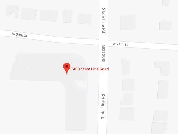 1. Eye Care and Specialty Surgery
- 7400 State Line Road
- Suites 100, 208 and 212
- Prairie Village, KS 66208
Office Contact: -
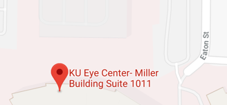 2. Medical Pavilion
- Eye Center
- 2000 Olathe Blvd., Miller Building, Suite 1011
- Kansas City, KS 66160
Office Contact:

Convenient payments with CommerceCare™
Commerce Bank has partnered with The University of Kansas Health System to bring you CommerceCare™, a fast, flexible and patient-friendly extended financing solution. With CommerceCare™, you can get many of the elective care services you want now and pay over time. CommerceCare™ offers two convenient plans so you can choose the option that works best for you.