A Life in Pictures

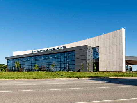
The new Quivira Ambulatory Surgery Center expands access to specialty surgical care.
Search our care options by diagnosis, treatment type or medical specialty.
Explore Specialties & Care
From common infections to rare diseases, we have the experts and leading-edge care.
Get guidance for scheduling an appointment, visiting a hospital, paying a bill and more.
Explore Patients & Visitors
Get the information you need to navigate your appointment or visit.
November 05, 2018
Expert neurosurgery puts life back in focus
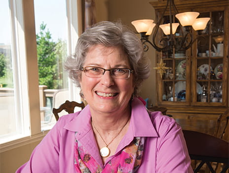
Kacey Hobson eagerly snapped more than 36,000 photos over the years to capture her big, extended family’s special milestones, from newborns to weddings to 90th birthdays. But Kacey was in front of the lens, not behind it, for before-and-after shots chronicling her own life-altering milestone. Side-by-side MRI scans show a tennis-ball-sized tumor pressing into the top of her brain in November 2013, but no trace of it in the June 2014 image. That happy day marked Kacey’s 6-month follow-up after complex brain surgery at The University of Kansas Health System.
A part-time nurse with energy to spare at 62, Kacey knew something was wrong in early 2013 when her usual stamina began to falter along with her balance. No firm diagnosis resulted from physician visits. By autumn, her left leg dragged slightly, her thinking was “fuzzy” and she was exhausted.
Scans revealed a meningioma – a benign, slow-growing tumor within the membrane covering her brain. While meningiomas are a relatively common brain tumor, the precarious placement of Kacey’s would make it tricky to remove. Risks included permanent paralysis on her left side or a major brain hemorrhage
From years as a pre- and post-op surgical nurse, Kacey knew she needed a top neurosurgeon. Contacting associates throughout the medical community to identify the region’s best specialists, she ultimately chose Paul Camarata, MD, neurosurgeon and neurosurgery program leader at The University of Kansas Health System.
"Dr. Camarata came in with solutions and immediately instilled confidence," she says. "I felt from the very beginning I had the right surgeon and the best neurosurgical team in the best facility in the area."
Dr. Camarata explains the challenges Kacey’s tumor posed. "It was underneath a large vein running along the top of the brain that carries a huge amount of blood, and compromising that could’ve been catastrophic," he says. "Also, her tumor lay adjacent to very important movement centers that control limb function. Put too much pressure on the brain during the procedure, and it can result in paralysis."
High-tech image-guided equipment helped Dr. Camarata and his team determine the tumor’s exact location. Performing a craniotomy, they opened Kacey’s skull by creating a 2-inch by 4-inch bone flap that functioned like a trap door. After eliminating the tumor, they replaced the flap, securing it with 2 ultra-thin, dime-sized titanium plates and teeny screws just under the scalp. The procedure spanned more than 5 hours.
"I was so impressed with and so thankful for my neurosurgical group, as well as every hospital staff member I encountered on my journey," Kacey said. "I loved the academic feel that permeated the entire experience."
She remembers little of the following week in intensive care. Another 9 days of inpatient rehabilitation improved her function and strength enough to continue recovery at home. Support from husband, Roger, and her family, as well as classes at Turning Point: The Center for Hope and Healing and The University of Kansas Health System’s brain tumor support group, have aided her emotional recovery.
Only a short time ago, Kacey’s future was highly uncertain. But today, this avid shutterbug confidently pictures the years to come – and can’t wait to see what develops.
We offer a variety of appointment types. Learn more or call 913-588-1227 to schedule now.