


- Home
- News Room
- Patient Stories
- First-Time Screening Mammogram

January 03, 2024
Megan Peters remembers caring for her father in his last days. She says it was a privilege and something she was honored to do, but it was also the hardest experience of her life.
“My dad had lung cancer, and I was there with him from his diagnosis until he passed away. I’m so glad I was able to do that, but I would do anything to ensure my children don’t have to go through the same thing – caring for a parent with advanced cancer.”
So, Megan got a mammogram.
Even though there is no history of breast cancer in her family, Megan, of Overland Park, Kansas, got her first screening mammogram shortly after her 40th birthday. The timing is in line with recent recommendations from the United States Preventive Services Task Force, an independent panel of national experts in disease prevention and evidence-based medicine. The panel updated its guidelines for breast cancer screening in May 2023 and now recommends mammograms starting at age 40 for all average-risk women.
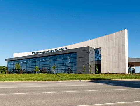
Megan chose The University of Kansas Hospital, Indian Creek Campus, for her mammogram appointment. There she received a 3D mammogram, which provides more detail than a traditional mammogram and is considered the most significant improvement in mammography in the past 30 years. The University of Kansas Health System uses 3D technology for all screening mammograms at every location.
Knowledge over nerves
Mammograms, especially the first one, can be anxiety-inducing for some women, but Megan knew she was in good hands. “From the time I walked in the door, everyone made me feel very comfortable and explained everything,” Megan says.
And she could be confident in the findings, knowing that her 3D mammography images were being read by dedicated, board-certified, fellowship-trained breast radiologists. Breast-dedicated radiologists at the health system read more than 31,000 breast images per year and find early-stage breast cancer at a rate that exceeds the national benchmark.
Finding breast cancer early is critical to achieving positive outcomes. Imaging that uncovers cancer in its earliest stages, when it is 98% curable, helps increase the likelihood that patients receive fast and minimally invasive treatments so they can return to their normal lives as soon as possible.
“I suppose every woman is a little nervous at first,” Megan says, “but I also know how important it is to detect any potential problems early. Having been through my dad’s experience, I want to make sure I’m screened for everything at the appropriate age and frequency. The knowledge about my health is more important than the initial nerves.”
Megan admits that she was a bit anxious about whether the mammogram would be uncomfortable. “I’d heard what other women said, and some told me their mammograms hurt, but my technologist was really great,” she says. “Before we did the actual screening, she explained how the machine works and what we would be doing. The mammogram itself only took a few minutes, and we were chatting the whole time.”
After being shown to a private changing room where she put on a soft, cotton top that allows for breast access, patients and their technologist enter a room with the mammography equipment. While the patient remains standing in front of the machine, the technologist positions one breast on a platform and explains where to place one’s arms and hands. The breast is gradually pressed against the platform by a clear plastic plate, flattening the tissue and holding it in place while taking the images. Each breast is positioned twice to take views from different angles, and the pressure is typically released after just a few seconds.
Megan Peters walks us through her first mammogram
Megan Peters:
I’m Megan Peters and I am at the Indian Creek Campus of The University of Kansas Health System to get my first mammogram. I just changed into my lovely robe and I am feeling a little nervous. I had some friends tell me that this is not necessarily a pleasant experience, but I’m ready to get it over with and get it off my to-do list and I’m waiting for the nurse to take me back to the exam room.
Emily:
And this is Mary Beth, my co-worker.
Speaker 3:
Hi! How are you?
Megan Peters:
I’m alright.
Emily:
Ok, come over here. I just want to go over a few things.
Megan Peters:
That was like, not that bad. I don’t understand why everybody’s freaking out about that, but maybe I just haven’t gotten to the hard part yet.
Emily:
Look over your right should for me, okay.
Megan Peters:
It was not that bad! I honestly was expecting it to be a lot more painful. It definitely was not pleasant, but it wasn’t painful at all, and Emily made it so easy. She really positioned me and I just kinda had to let her do the movements, and in and out pretty quickly.
Emily:
Alright, we are all finished, you did awesome.
Megan Peters:
Thank you.
Emily:
We’re going to head to the right here.
Megan Peters:
Okay.
So now it feels like the hard part is over, but now I have to wait for my results. Emily, my nurse, told me I should be getting my results later today via MyChart, which is so great, because I didn’t really think past getting the actual mammogram and the nervousness that might kind of come in afterwards waiting for the results, so being able to know fairly quickly is such a wonderful gift and I’m excited to get those results later today. Hopefully they will be all good.
Making a commitment to health
In addition to the benefit of expert eyes reviewing Megan’s images, mammograms at the health system use 3D technology. Multiple views of the breast tissue combine to create a 3D image. This type of mammography reduces the need for additional imaging in many patients and also detects more cancers than 2D mammography. 3D mammograms are the preferred type of imaging for all patients, no matter the breast density.
“I was done before I knew it and able to go about my day,” Megan says. Results are usually available in the patient’s online medical records portal within 24 hours. In Megan’s case, results appeared only a few hours after her exam – and she received the reassuring news that her mammogram showed no evidence of cancer.
If the radiologist does detect a potential abnormality, the patient is asked to return for additional imaging. Patients at high risk may undergo routine breast MRI, ultrasound or contrast enhanced mammography in addition to an annual screening mammogram. It’s important for each individual to discuss their own personal breast screening recommendations with their primary care physician.
“We know mammograms are really important, and since I’m committed to taking care of myself, these screenings are just going to be part of my annual routine from now on,” Megan says. “Now I have a good baseline going forward, and I feel more relaxed knowing I’ve done what I need to do to ensure I’ll be around for my kids for a long, long time.”

Schedule your mammogram online
We find early-stage breast cancer at a rate that exceeds the national benchmark.